Cortisol & Menopause: What Your Levels Reveal
Research shows cortisol levels during the menopausal transition are driven by estrone glucuronide, FSH, and testosterone — not stress alone. Here's what the dat


If you've been waking up exhausted, running hot at night, and feeling like your body has shifted into a strange new gear, you're not imagining it. Research from the Seattle Midlife Women's Health Study confirms that cortisol levels during the menopausal transition rise in measurable, biologically driven ways — and the drivers are not quite what most people expect. Understanding cortisol levels during the menopausal transition may be one of the most important pieces of your midlife health picture.
The study tracked 132 women across the late reproductive stage, early and late menopausal transition (MT), and early postmenopause — collecting more than 5,000 overnight urine specimens over 15 years. What emerged is a detailed, evidence-based view of how the stress hormone cortisol behaves as the ovaries begin to wind down.

Why Cortisol Rises During the Menopausal Transition
Cortisol is the body's primary stress-mobilising hormone, produced by the adrenal glands in response to signals from the hypothalamic-pituitary-adrenal (HPA) axis. From a woman's thirties onward, baseline cortisol climbs gradually — a well-documented effect of ageing. But the menopausal transition adds a distinct biological layer on top of that background rise.
The HPA axis and the hypothalamic-pituitary-ovarian (HPO) axis are deeply interconnected. As HPO function shifts during the MT — with oestrogen fluctuating, FSH climbing, and the menstrual cycle becoming irregular — the HPA axis responds in kind. This is not simply a stress reaction. It is an integrated hormonal recalibration.
The late MT stage is particularly significant. Menstrual cycles exceed 60 days, FSH surges, and hot flashes increase in frequency and severity. The Seattle study found that overnight cortisol levels rose measurably as women moved from the early into the late MT stage — a finding that points squarely at reproductive hormone shifts, not lifestyle stress, as the primary trigger.
The Hormones Most Strongly Linked to Cortisol
Three reproductive hormones stood out as the strongest predictors of overnight cortisol levels in multivariate analyses: estrone glucuronide (E1G), FSH, and testosterone. Each was independently and significantly associated with elevated overnight cortisol (p<.0001). Together, they formed the best predictive set in the entire model.
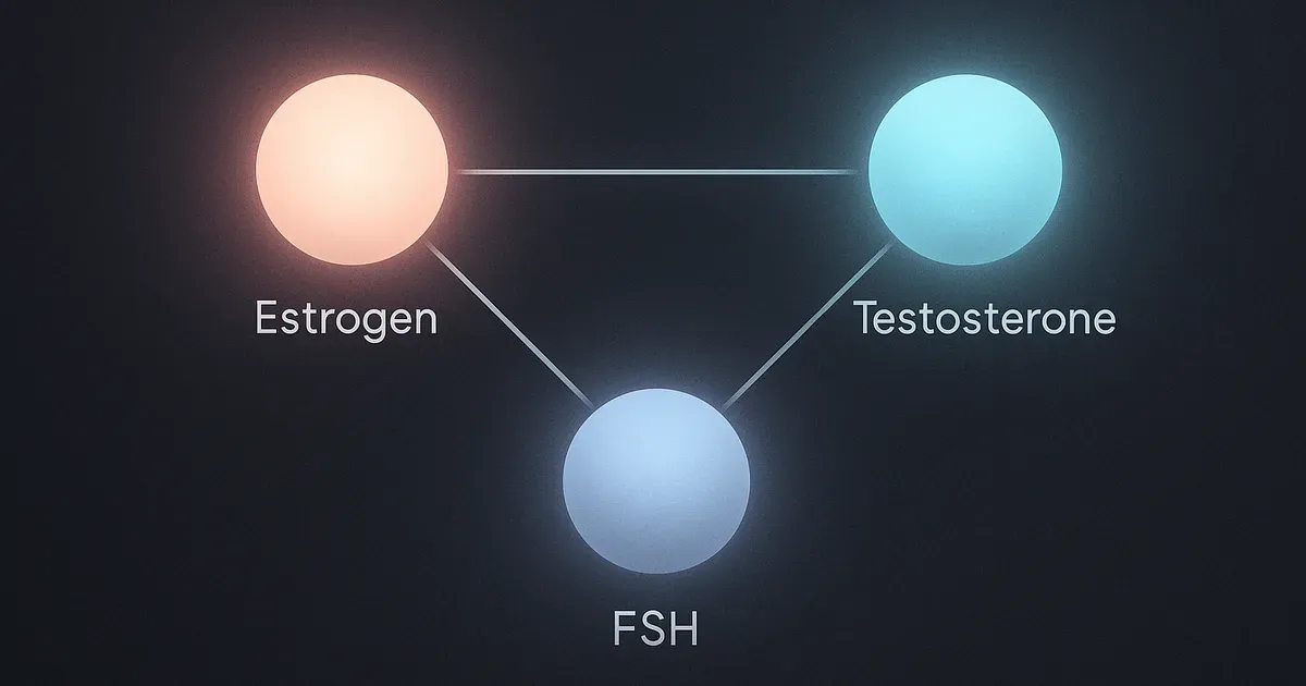
Estrone glucuronide is a urinary metabolite of oestrone, the predominant oestrogen in postmenopausal women. Higher E1G was linked to higher cortisol, which aligns with earlier laboratory findings showing that both endogenous and exogenous oestrogen elevate cortisol. This challenges the assumption that falling oestrogen is the sole hormonal story of menopause — the picture is far more dynamic.
FSH, or follicle-stimulating hormone, rises as the ovaries become less responsive to signalling. Its strong association with cortisol suggests that the pituitary's increased drive to stimulate the ovaries may simultaneously influence adrenal output. Testosterone, often overlooked in discussions of women's hormonal health, was also a meaningful predictor — underlining the breadth of hormonal interplay involved.
The Role of Stress Hormones: Epinephrine and Norepinephrine
Beyond reproductive hormones, the catecholamines — epinephrine and norepinephrine — were each significantly associated with overnight cortisol levels (p<.0001). These are the fight-or-flight neurotransmitters released by the adrenal medulla during acute and sustained stress. Their correlation with cortisol confirms that the sympathetic nervous system and the HPA axis are working in concert during the MT.
This is relevant for women who experience heightened anxiety, heart palpitations, or a general sense of being on edge during perimenopause. Those experiences may have a measurable neurochemical basis — not just an emotional one.
What the study did not find is equally important. Perceived stress, mood symptoms, sleep disturbance, memory complaints, and social factors such as income, role burden, employment, and social support had little independent relationship to overnight cortisol once the biological variables were accounted for. The biology appeared to dominate.

What Does Not Drive Cortisol Levels as Much as Expected
Many women — and many clinicians — assume that life stress is the main reason cortisol climbs during midlife. The Seattle data complicates that assumption significantly. Factors including perceived stress, history of sexual abuse, parenting demands, and social support were tested rigorously. When reproductive hormone levels were included in the model, these social and psychological variables lost their predictive power.
This does not mean stress is irrelevant to women's health during the MT. It means that when it comes to overnight cortisol specifically, the hormonal environment of the menopausal transition appears to be the dominant driver.
Similarly, depressed mood, BMI, perceived health, and smoking — all factors associated with cortisol changes in other populations — did not emerge as significant independent predictors in this dataset once E1G, FSH, and testosterone were in the model. The biological signal was simply stronger.
Hot flashes deserve a separate note. Early laboratory studies had shown cortisol spikes coinciding with hot flash events measured via sternal skin conductance. The Seattle study's earlier reporting also linked more severe hot flashes with higher cortisol. But in multivariate analyses, hot flash severity as a standalone symptom was not the primary explanatory variable — reproductive hormone levels were.
Cortisol, Metabolic Health, and Long-Term Risk
The implications of rising cortisol during the MT extend well beyond sleep and mood. Sustained cortisol elevation has downstream effects on metabolic function, bone density, cognitive performance, and cardiovascular risk. Women in the late MT stage already show higher total cholesterol, elevated LDL-C, higher apolipoprotein B, and greater VLDL cholesterol — all markers of increased metabolic risk.

Chronically elevated cortisol is associated with lower bone density in older women, and with minor cognitive complaints — the kind of memory lapses that women often attribute to "brain fog" during perimenopause. If rising cortisol is a biological feature of the MT rather than a stress response, this reframes how clinicians and researchers should think about these complaints.
The relationship between rising cortisol and metabolic syndrome is also worth tracking closely. If the HPA axis upregulation during the MT contributes to insulin resistance, abdominal fat deposition, or dyslipidaemia, then cortisol levels during this window could serve as an early biological marker of longer-term cardiometabolic risk. This is an area where future research is critically needed.
Hormones do not act in isolation — the interplay between adrenal output, reproductive hormones, and metabolic signalling during the MT is a system-level phenomenon, not a single-gland story.
Understanding the Study's Scope and Limitations
The Seattle Midlife Women's Health Study is unusually rigorous for research in this area. Using overnight urine specimens — rather than single daytime blood draws — captures integrated cortisol output over hours rather than a snapshot. With up to 5,218 observations across 132 women spanning 15 years, the dataset has the statistical depth to detect real effects and rule out noise.
Multilevel modelling was used to handle the nested structure of the data — repeated observations within women across time — which appropriately accounts for individual differences in cortisol baselines. Women using hormone therapy or corticosteroids were excluded, removing two major confounders.
That said, the sample was drawn from a specific demographic: predominantly educated, employed, and with moderate-to-higher household incomes. Generalisability to women with different socioeconomic backgrounds, ethnicities, or health profiles warrants careful consideration. The study also did not assess adrenal androgens like DHEA-S in relation to cortisol within this analytical framework, which could add further nuance.

The Bottom Line
Cortisol levels during the menopausal transition are driven primarily by reproductive hormone changes — specifically by shifts in estrone glucuronide, FSH, and testosterone — rather than by psychological stress, lifestyle habits, or social circumstances alone. The HPA and HPO axes are in constant dialogue, and when one shifts, the other responds.
For women navigating perimenopause and the challenges it brings — disrupted sleep, hot flashes, mood shifts, memory concerns — this research provides an important reframe. Many of these experiences may have a measurable, biological hormonal basis. They are not simply the result of life getting harder or stress levels climbing.
For clinicians, the data suggest that overnight cortisol measurement could serve as a meaningful biomarker during the MT, and that reproductive hormone status should be central to any interpretation of elevated cortisol in midlife women. Addressing the hormonal environment, rather than focusing narrowly on stress reduction, may be the more productive clinical direction.
The broader picture connects cortisol to long-term risks in bone health, cognition, and cardiovascular disease — making the menopausal transition not just a reproductive event, but a critical window for whole-body metabolic monitoring.